Seeing our life pass us by is something we often don’t think about until our vision changes. We often don’t think about how our eyes work or what goes in to viewing an image daily. Here are the basics of the eye.
An Eye Outline:
On average a person’s eye is about 1 inch in diameter. There are two areas “The Front Portion” and “Behind the Lens”. The front side has the parts that control or include lighting an image or refracting light, and most of this portion is what we see when we look at a person
Front Portion of the Eye:
Iris – Is the pigmented area
Cornea – The cornea is a clear dome over the iris.
The Pupil – Black circle in the center of the eye that refracts the light to the back of the eye.
The Sclera – The white area of the eye
The Conjuctiva – a layer of tissue that covers the front of the eye.
The lens – Focuses the light toward the back of the eye to help develop the image.
Behind the Lens:
While you can not visible see what goes on behind the lens and many might think it looks rather plain behind the lens, there are several things going on within the eyeball. Below you will find more parts located within the eyeball behind the lens.
Vitreous – This is the gel which fills the eye
Retina – Light sensitive cells in the lining of the eye which converts the light into electrical impulses.
Optic Nerve- The optic nerve is located behind the eye and carries the electrical impulses to the brain to help form an image
Macula – The macula is a small extra sensitive area in the retina which controls central vision
Although this is only a preview of the eye it can help give you a better idea of some of the working parts within your eyeball. In combination these parts help you view the images your eyes see every day.
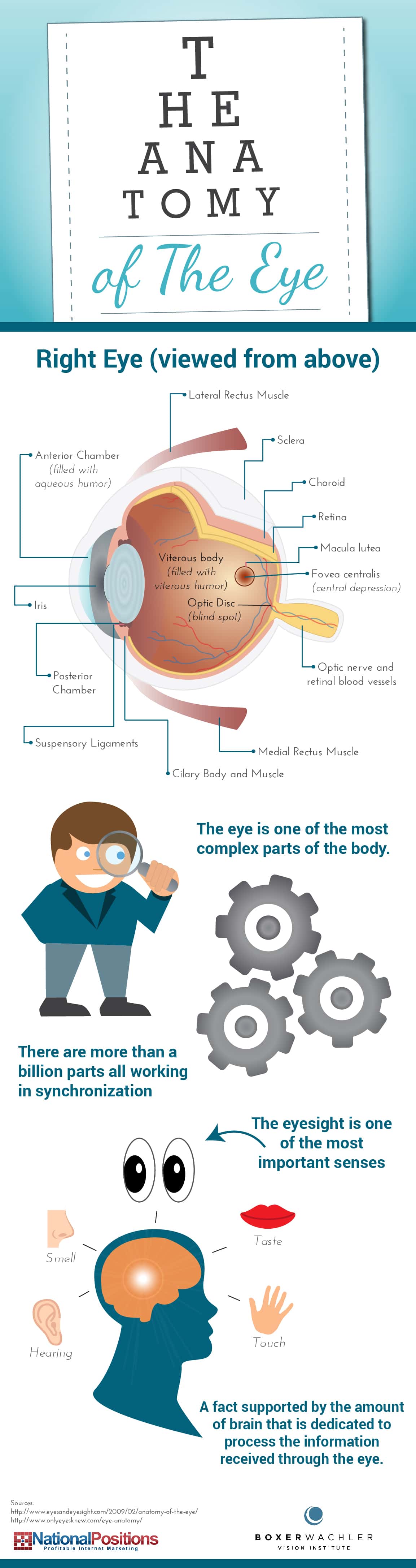
Here is an interesting graphic about the Eye Anatomy: (click on the image to see a larger version)